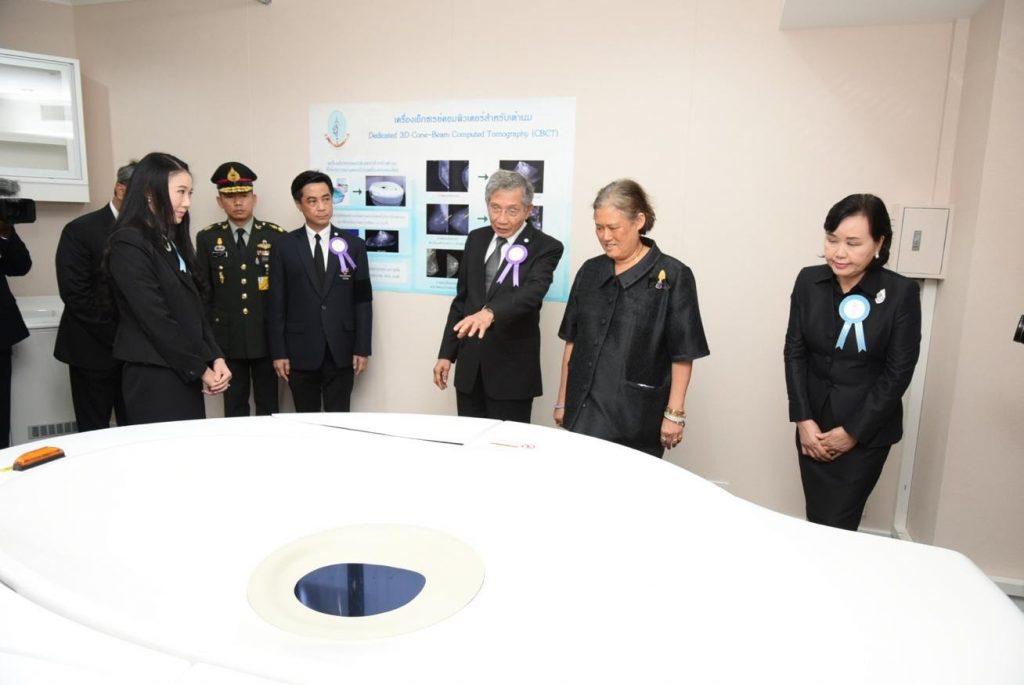
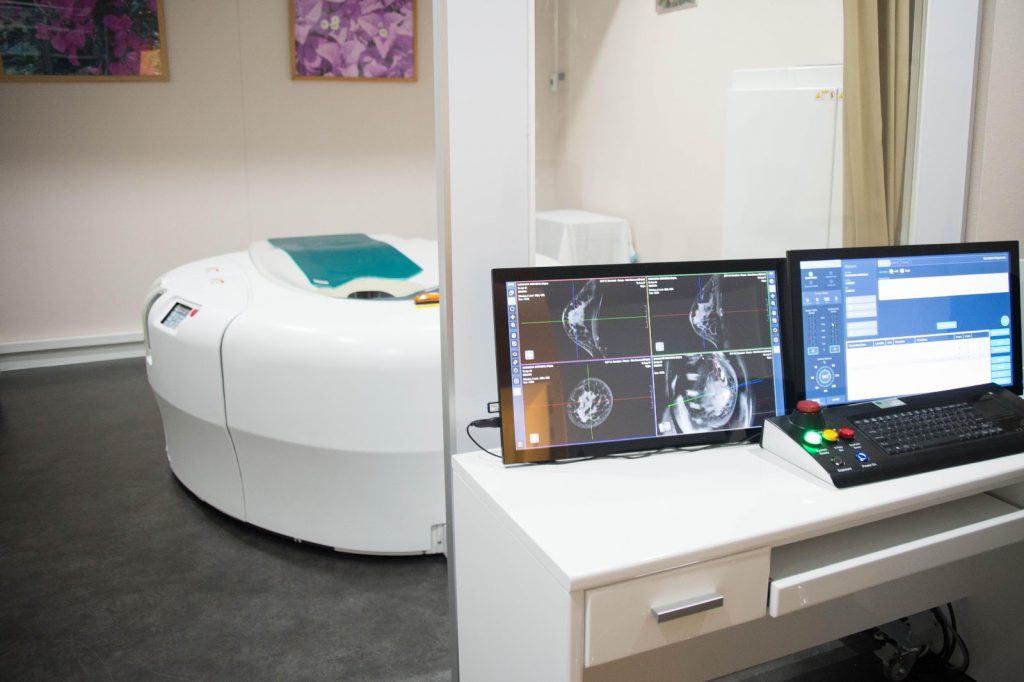
In March 2017, H.R.H. Princess Maha Chakri Sirindhorn visited the new CT-mammography machine, at the Queen Sirikit Centre for Breast Cancer, the latest addition to the battery of equipment for the diagnosis and treatment of breast cancer at the QSCBC. The CT-mammography is the first complete unit in the world, which provides a true 3-D picture of the breast without compression. The extra clarity and complexity of the image supplements the information from the standard mammography and can greatly help in the diagnosis and the planning of breast conservation surgery.
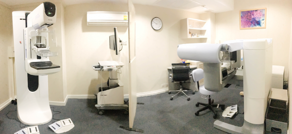
Breast Radiology Unit
CT Mammography
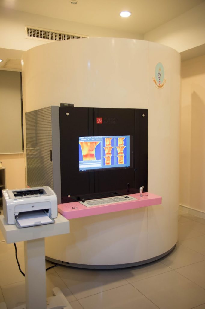
Thermography
The addition of the latest ‘thermography’ equipment measures differences of infrared emissions from the breast and can help to pinpoint lesions of abnormality. Thermography gives out zero levels of radiation, which has been a controversial issue for patients and can be repeated as often as required. Some women are discouraged from having annual breast cancer screening due to the fear of pain and discomfort, but the CT- Mammography and Thermography do not require breast compression during the examination. Thermography is very useful for younger patients
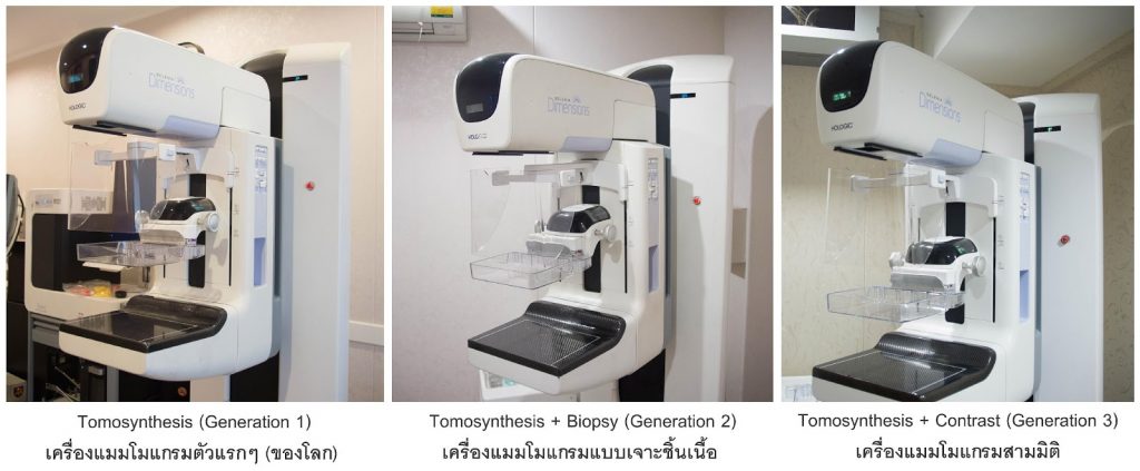
Digital 3-D Tomosynthesis
The QSCBC has three tomosynthesis digital mammography machines (3D), which are used as a standard mammography examination.
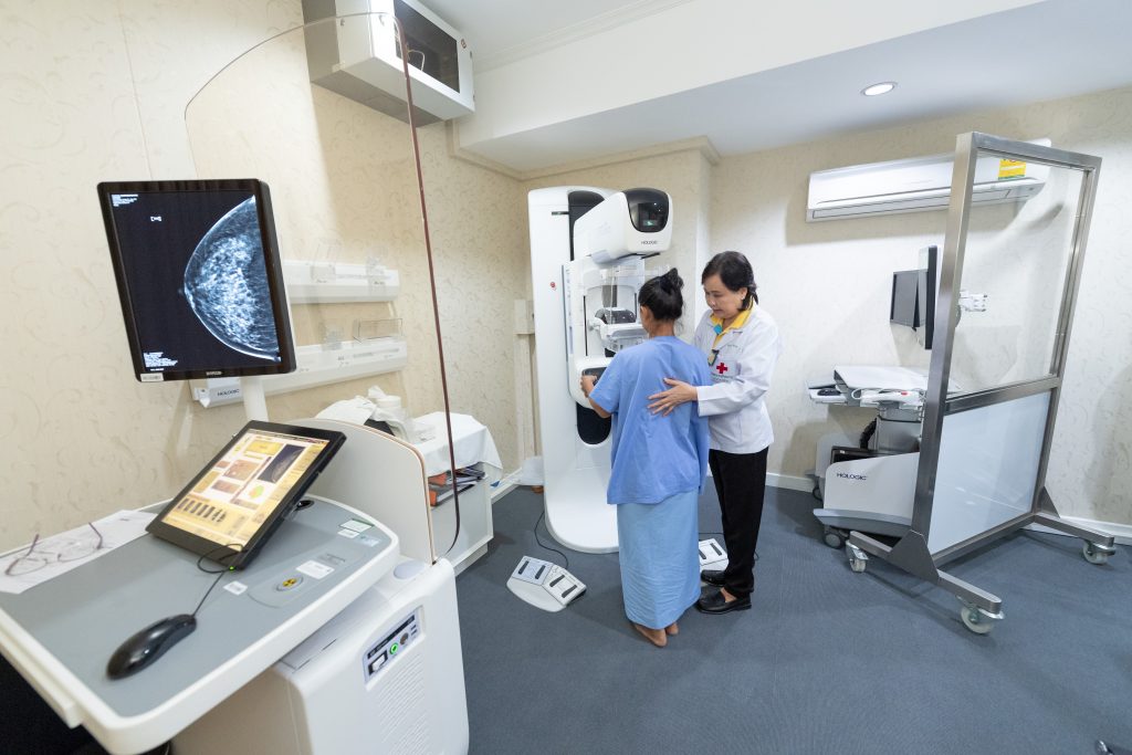
Mammogram uses X-rays to capture breast images. However, in the case that a patient has a high risk of breast cancer, the QSCBC will use Dedicated Breast MRI, CT Scan and ultrasound investigations to confirm a complicated and difficult diagnosis. Radiation emitted from a modern digital mammography machine is now significantly lower and considered safe, if given in approved regular intervals.
The patient places one breast at a time on a small ledge and a compression plate is lowered gently onto the breast compressing the whole area. The mammogram will take pictures from various angles. ( 2d OR 3 D AS required.). The breast is compressed for a few seconds while the machine is working and the patient holds their breath during this very brief period.
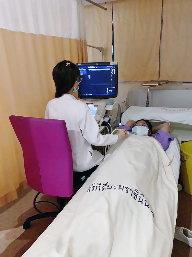
Breast Ultrasonography Examination
Breast Ultrasonography Examination is a radiological examination which uses high frequency sound waves to generate images inside the breast. It is very useful to supplement the tomosynthesis mammography used at the QSCBC, to detect structural abnormalities, e.g. the difference between a fluid filled cyst and a solid tumour, which will help give an accurate diagnosis. It does not, however, detect calcification in the breast as well as mammography. The two supplement one another and are used routinely together, for the last 25 years. The QSCBC introduced this approach as routine.
The ultrasound does not use radiation and is harmless.
The patient lies down on the bed, then a radiologist applies gel on the breasts to help the sound wave travel through the skin more easily. The patient can see the captured images on the monitoring screen while having an examination if they wish to do so. Needle guide biopsies also use ultrasounds to pinpoint the exact position of possibly abnormal lesions in the breast under local anaesthesia.
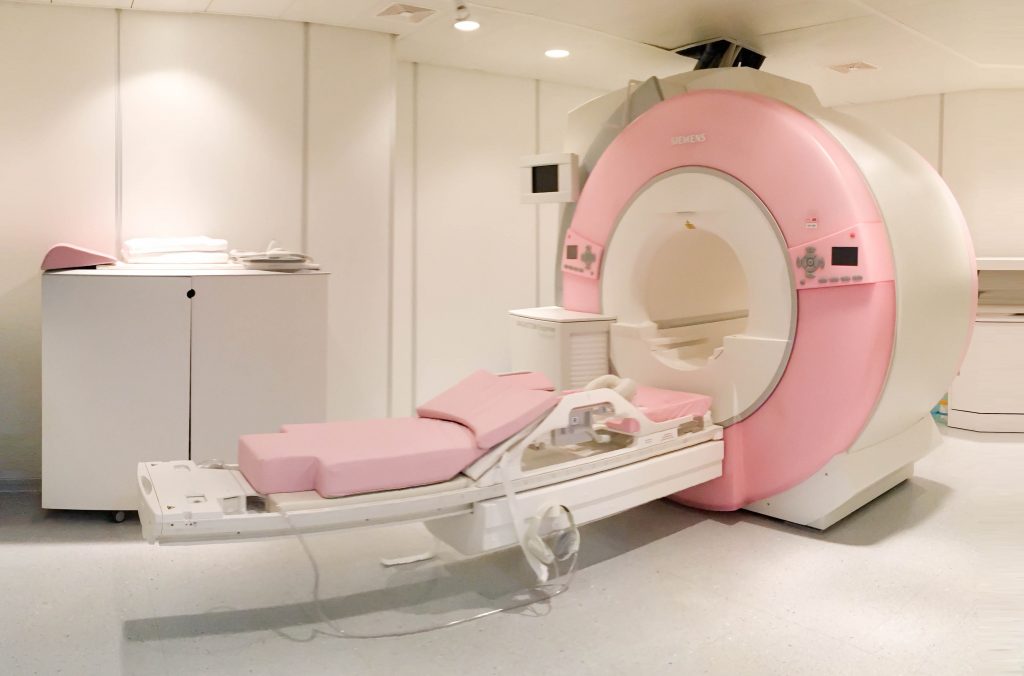
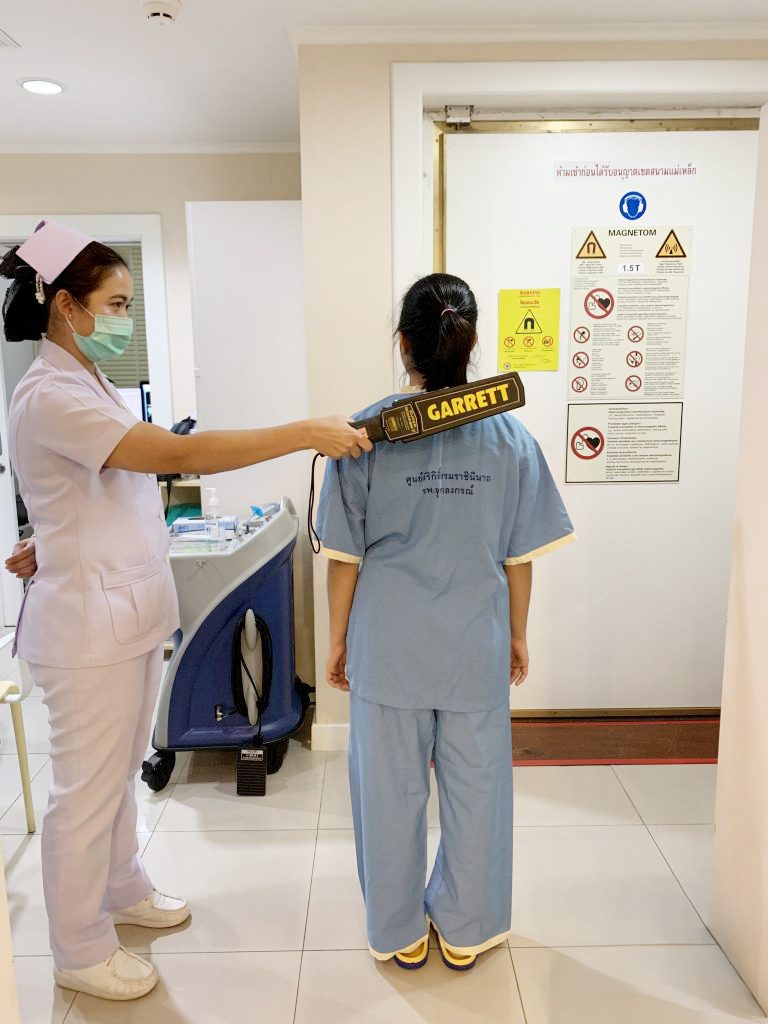
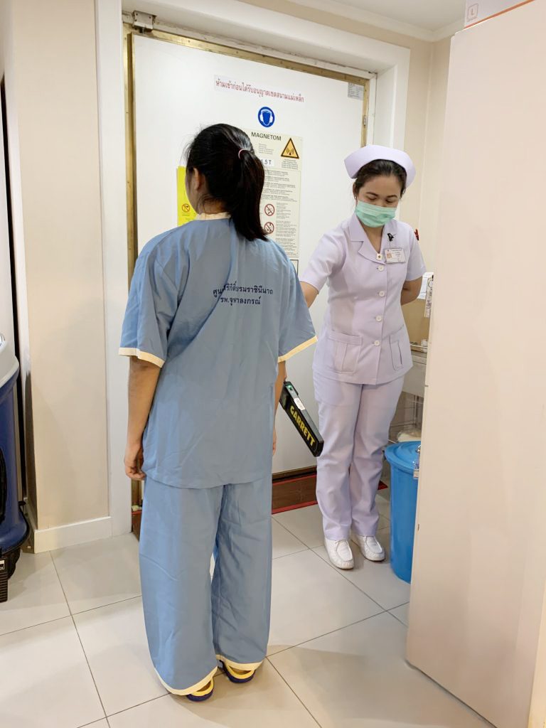
Dedicated breast MRI Examination
Dedicated Breast MRI is a radiological examination using electromagnetic waves. It can help to detect the exact position and type of lesions more precisely, it supplements other diagnostic examinations , like mammograms and ultrasounds. It helps to monitor function eg the blood flow in the breast, as well as, structure of the breast in question. It is particularly useful with patients who already have breast prostheses. It does, however, require the patient to be injected intravenously with a type of very low grade radioactive dye and the patient needs to be checked for allergies and kidney function.
The examination takes only 30 minutes. The patient lies face down on a sliding bed which slowly moves into the MRI machine, which is like a large ring, but not a tunnel, so the patient will not be claustrophobic. This type of examination requires no breast compression.
The Queen Sirikit Centre for Breast Cancer (QSCBC) is the first hospital in Thailand to use a Dedicated MRI scanner to examine for breast cancer ,( 6-8 patients can be examined per day.) The Queen Sirikit Centre for Breast Cancer also developed the image analysis from the Dedicated Breast MRI, in order to detect breast defects more clearly and faster.
Currently, people with a high risk of breast cancer are advised to be examined with an MRI; People with an average risk of breast cancer, do not need to do so, but the MRI examination can help to prepare a patient for surgery, by for example, doing needle guide biopsies which can locate the position of a lesion more accurately.
The number of patients who receive the MRI breast examination at the QSCBC is 70 – 90 cases per month.